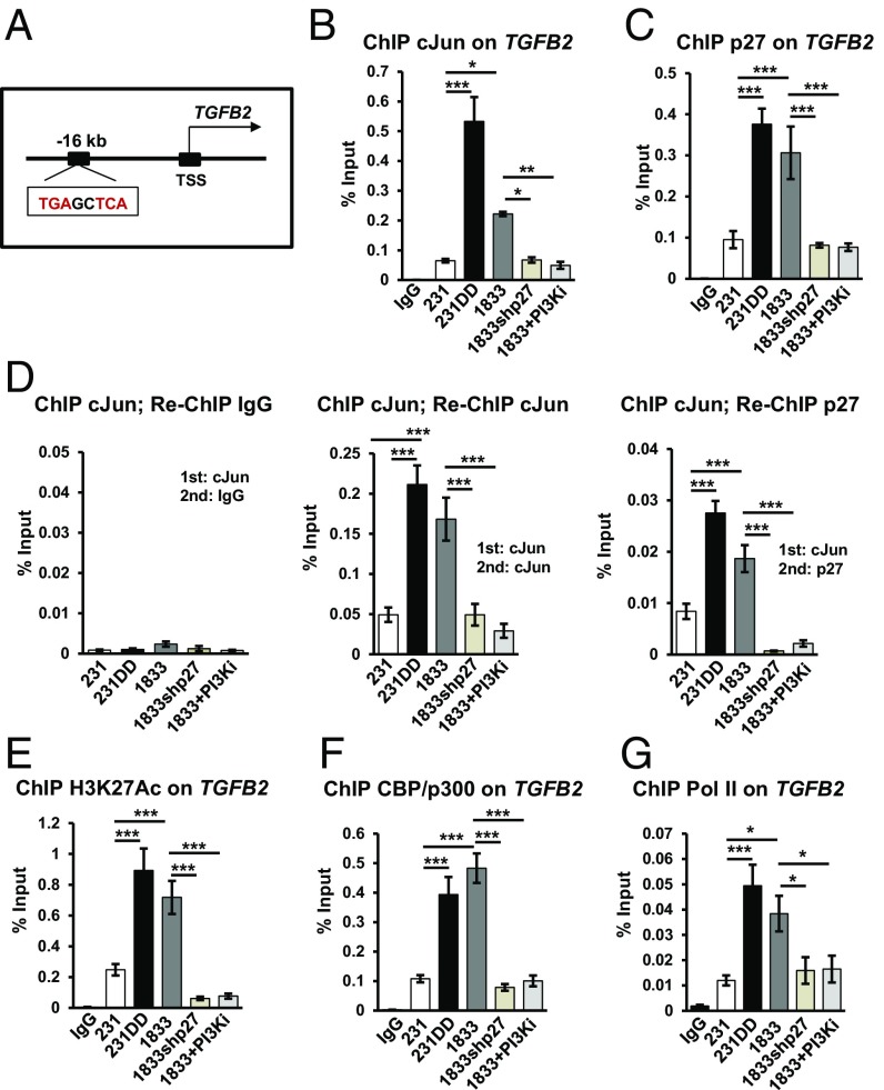
Fig. 4.
cJun and p27 are corecruited to a TGFB2 AP-1 site. (A) cJun/AP-1 consensus motif upstream of TGFB2 TSS. (B and C) ChIP-qPCR with cJun (B) and p27 (C) antibodies show binding to an AP-1 site upstream of TGFB2 in the indicated lines and in 1833 treated for 48 h with PF1502 (PI3Ki). (D) ChIP-re-ChIP assay of p27 association with cJun-bound TGFB2 AP-1 site in the indicated lines. (E–G) ChIP assays show H3K27Ac (E), CBP/p300 (F), and Pol II (G) binding to the TGFB2 AP-1 site in the indicated lines. Means ± SEM graphed from three or more replicates of three or more biologic assays; post hoc P values from ANOVA (*P < 0.05, **P < 0.01, ***P < 0.001). See also SI Appendix, Fig. S3. 231DD, 231p27CK−DD.

