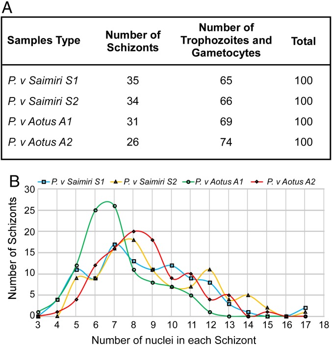
Fig. 1.
P. vivax Sal I infection in Saimiri and Aotus monkeys. (A) Two Saimiri monkeys and two Aotus monkeys were infected with the P. vivax Sal I parasites. Late schizont stage parasites were purified from monkey blood samples for RNAseq analysis. Following enrichment by culture, a Giemsa smear was performed on two Saimiri and two Aotus infections to evaluate the percentage of schizonts and the stage of schizont development. The table summarizes the proportion of schizont-stage parasites in a total of 100 parasites for all infections studied. (B) Graph shows the number of nuclei in each schizont in two Saimiri and two Aotus P. vivax samples. A total of 100 schizonts were counted for the total number of nuclei per schizont for each sample.

