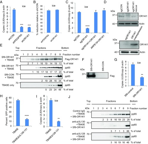
Fig. 3.
OR14I1 is necessary for PC+ HCMV binding to and infection of epithelial cells. (A) Virus binding assay. ARPE-19 cells expressing the indicated shRNAs were incubated with TB40E-GFP virus on ice (MOI 3.0). After washing, cell surface-bound viral DNA (UL83) was quantified using qPCR and normalized to cellular DNA (β-actin). (B) The results in A are presented as the relative reduction of viral DNA in the knockdown cell lines relative to shCON. (C) Binding assays as in A using ARPE-19 cells expressing the indicated sgRNAs and/or cDNAs: sgCON, clonal sgOR14I1 cells, sgOR14I1 cells expressing sgRNA-resistant OR14I1, or WT cells overexpressing OR14I1 (MOI 3.0). (D) Immunoblots of cells in C. (E) Membrane flotation assay. TB40E-GFP virus was incubated with membrane vesicles from control Sf9 cells (Sf9-CON) or Sf9 cells expressing human Flag-OR14I1 (Sf9-OR14I1). After centrifugation, fractions underwent immunoblotting to determine the levels of TB40E-GFP virus (virion protein pp65) and location of membrane vesicles (Flag-OR14I1). (F) Immunoblot of cells in E. (G) TB40E-GFP virus was preincubated with Sf9-control or Sf9-Flag-OR14I1 membrane vesicles before being used in a virus binding assay with ARPE-19 cells (MOI 3.0). Viral (UL83) and cellular (β-actin) DNA levels were quantified by qPCR. (H) ARPE-19 cells infected with TB40E-GFP or a PC-deleted TB40E-GFP (TB40E∆UL128–131; MOI 3.0). Cells were fixed at 2 dpi and assessed for GFP (green)-positive cells. (I) ARPE-19 cells were incubated with either TB40E-GFP or TB40E∆UL128–131 virus (MOI 3.0) on ice. After washing, cell surface-bound viral DNA (UL83) was quantified using qPCR and normalized to cellular DNA (β-actin). (J) Control IgG antibody, anti-pUL128, or anti-pUL130 was preincubated with purified TB40E-GFP virus in a membrane flotation assay. Data represent the mean of n = 3 experiments ±SD. **P < 0.01, ***P < 0.001, ****P < 0.0001.

