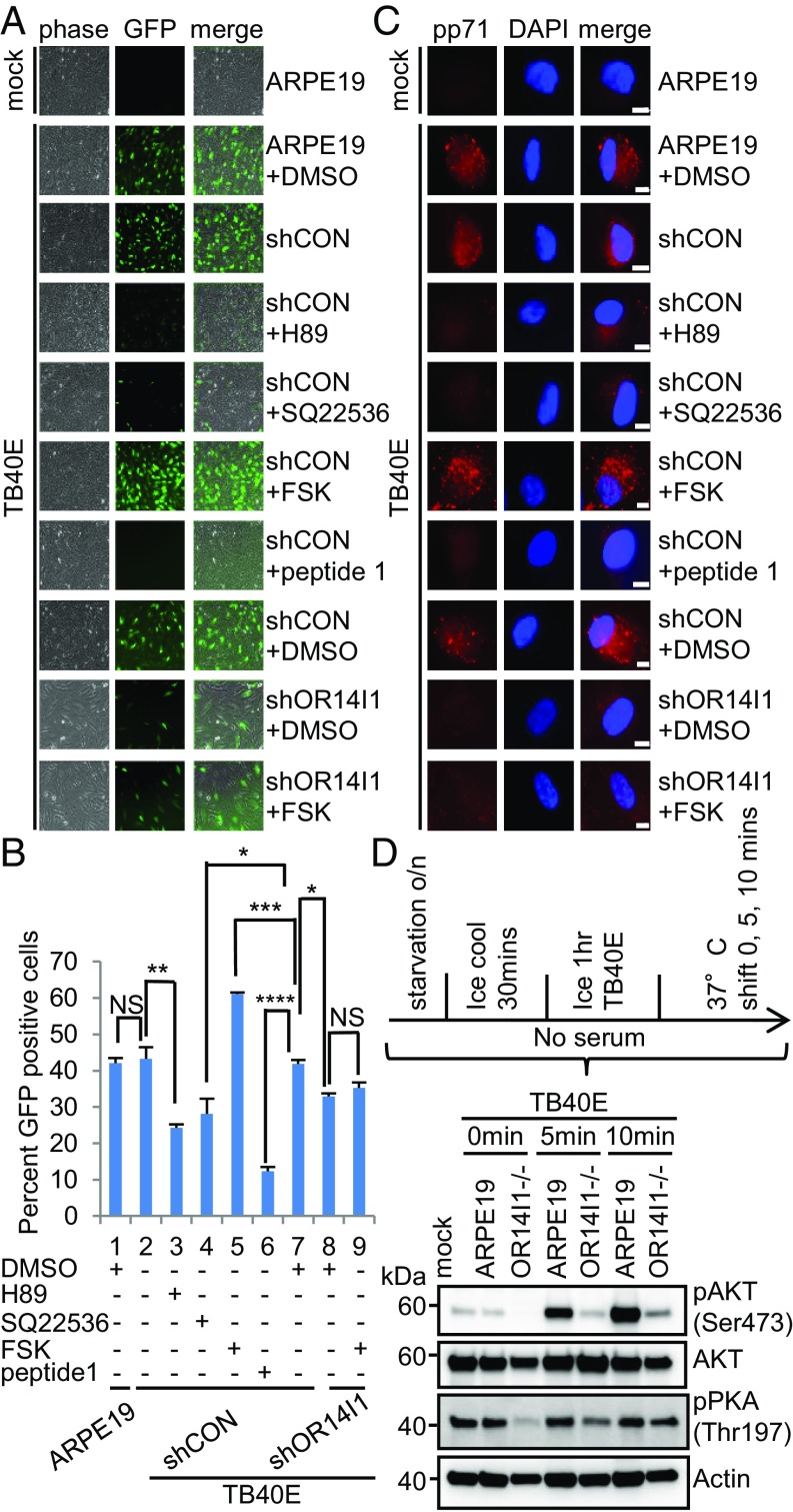
Fig. 6.
AC/PKA/AKT signaling is required for HCMV entry and infection of epithelial cells. (A) ARPE-19 cells expressing the indicated shRNAs were pretreated with PKA inhibitor H-89 (20 μM), adenylate cyclase antagonist SQ22536 (150 μM), AC agonist forskolin (20 μM), or DMSO solvent for 2 h before TB40E-GFP infection (MOI 2.0). Peptide 1 was preincubated with TB40E for 2 h at 37 °C before addition to cells. Cells were imaged (10×) for GFP at 2 dpi. (B) Quantification of data in A after cell fixation and DNA staining. Results are presented as the percent GFP-positive cells. Data represent the mean of n = 3 experiments ±SD. *P < 0.05, **P < 0.01, ***P < 0.001, ****P < 0.0001. (C) ARPE-19 cells expressing the indicated shRNAs were pretreated with the indicated drug for 2 h before TB40E-GFP infection (MOI 2.0); peptide 1 was preincubated with TB40E for 2 h at 37 °C. Cells were cooled on ice for 30 min and then infected with cold TB40E (MOI 2.0) containing the noted small molecules for 1 h on ice. Cells were then transferred to 37 °C. At 2 hpi, cells were washed and treated briefly with trypsin to remove surface-bound virus. They were then fixed, permeabilized, stained with anti-pp71, and imaged (63×). (Scale bars, 10 µm.) Representative images of three independent experiments are shown. (D) ARPE-19 and OR14I1−/− cells were infected with cold TB40E (MOI 2.0) for 1 h on ice. Cells were transferred to 37 °C for 0, 5, and 10 min. Levels of p-AKT (S473), total AKT, p-PKA (T197), and actin were detected by immunoblotting from whole-cell lysates. o/n, overnight.

