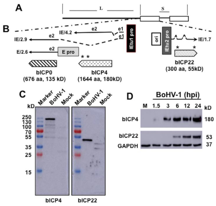
Figure 1: Schematic of IE region and location of the bICP0, bICP4, and bICP22 genes.
Panel A: Structure of BoHV-1 genome and location of unique long (L) region, direct repeats (open rectangles), and unique long region (S).
Panel B: The IE/4.2 mRNA encodes the bICP4 protein and IE/2.9 mRNA encodes the bICP0 protein. A single IE promoter activates expression of IE/4.2 and IE/2.9 (IEtu1; black rectangle). E/2.6 is the early bICP0 mRNA and is regulated by the bICP0 early promoter (E pro; gray rectangle). bICP0 protein coding sequences are in Exon 2 (e2). Origin of replication (ORI) separates IEtu1 from IEtu2. IEtu2 promoter (IEtu2 pro) regulates IE1.7 mRNA expression, which is translated into the bICP22 protein. Solid lines in IE/2.9, IE/4.2, and IE/1.7 are exons (e1, e2, or e3) and dashed lines introns. Location and size of the three proteins encoded by IEtu1 and IEtu2 are shown: arrows denote directionality of the proteins. The bICP4 peptides synthesized for producing the peptide specific antibody are: amino acids 1461–1478 (RRAGQAPGREAREGRGRG) and amino acids 1604–1621 (GVSPWGSRGVRAFRRPPG). The peptides synthesized for producing the bICP22 antibody are: amino acids 26–40 (GPAPADEHARRGPGA) and amino acids 281–296 (GSPSGRARARPAPAKR). An asterisk denotes the location of the peptides within the bICP4 and bICP22 ORFs.
Panel C: Monolayers of CRIB cells were mock infected or infected with BoHV-1 (MOI=2) and whole-cell lysate prepared at 12 hours after infection. Proteins (50 ug protein in each lane) were separated in a 10% SDS-PAGE and detected by Western blotting using the rabbit anti-bICP4 or bICP22 peptide antibody as described in Materials and Methods. The marker lane is a Thermo Scientific Page Ruler pre-stained protein marker and the size of these proteins is denoted on the left.
Panel D: As described above, CRIB cells were mock infected (lane M) or infected with BoHV-1 (MOI=2) for the denoted time (hours after infection). Whole-cell lysate (50 ug protein in each lane) was separated in a 10% SDS-PAGE and detected by Western blotting using the rabbit anti-bICP4 or bICP22 peptide antibody. GAPDH protein levels were analyzed in the respective samples as a loading control. Approximate size of the respective proteins is denoted on the right of the Western Blots.

