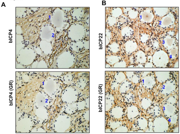
Figure 5. Identification of GR+ TG neurons that express bICP4 or bICP2 in the same TG neuron.
IHC was performed using the bICP4 or bICP22 antibodies as described in Figures 2 and 3 using sections cut from TG of latently infected calves at 6 h after DEX treatment to initiate reactivation from latency. IHC was also performed using a GR-specific antibody on consecutive sections. Numbers denote TG neurons that were bICP4+ or bICP22+ and GR+.

