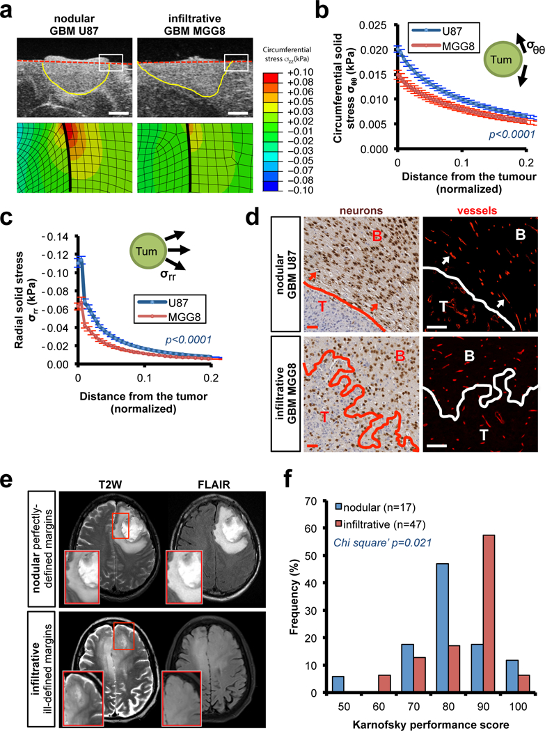
Fig. 1. Measuring how nodular tumors mechanically affect the surrounding brain.

(a-c) Solid stress in mouse models of nodular (patient-derived U87 cell line) and infiltrative brain tumors (patient-derived MGG8 cell line) in nude mice. (a) High-resolution ultrasound imaging of the stress-induced deformation and representative stress profiles across the tumour diameter and the normal surrounding tissue. Scale bar: 1 mm. (b-c) Estimation of the circumferential (compression; σθθ) and radial (tension; σrr) stress in the surrounding tissue obtained from mathematical model (described in Figure S1b-d and Method section). Data are mean of three independent tumors ± s.e.m. (d) Micro-anatomic deformation of the brain tissue around nodular tumors. IHC of U87 and MGG8 tumors at the interface of the normal brain tissue. Neurons (NeuN staining) and vessels (Collagen IV staining). Arrows indicate the deformed region around the tumour, neurons are packed and vessels are displaced following the circumference of the tumour margin. Representative images from a cohort of 10 mice with tumour. Scale bar: 50 μm. (e) Representative T2W and FLAIR MRI of pre-surgery pre-treatment patients with perfectly-defined margins GBM (nodular) and ill-defined GBM (infiltrative). (f) Karnofsky performance score (KPS) histograms of patients with perfectly-defined margins (nodular) or ill-defined GBM (infiltrative). Cohort of 64 pre-surgery pre-treatment GBM patients.
