
Fig. 6. Decompression restores vessel perfusion and partially rescues motor-coordination.
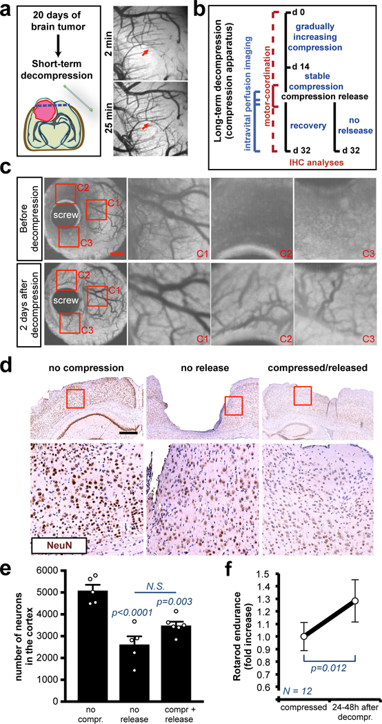
(a) OCT angiography after decompression of brains with nodular U87 tumors by removing cranial window (craniotomy). Complete time-course in Figure S9B-C. (b) Schematic of the long-term compression/decompression timeline via the in vivo chronic apparatus. (c) Longitudinal OCT angiography before and after decompression. Images of a representative mouse from a cohort of 3 mice. Insets are magnifications of the red squares. See Movie S7 for the complete time-lapse. Scale bar: 1 mm. (d) Representative cortexes stained with anti-NeuN from a cohort of 3 mice. The long-term compression induces tissue loss and the decompression partly restores the micro-anatomy of the cortex. Insets are magnifications of the red squares. Scale bar: 500 μm. (e) Quantification of the NeuN+ neurons in the cortexes, decompressed or compressed and released. Data are mean ± s.e.m. (f) Rotarod endurance (index of motor coordination and balance) 24–48 hours after decompression of the motor and somatosensory cortex. Data are mean of 2 consecutive days and 12 mice per group ± s.e.m (normalized with the value before decompression).