
Fig. 7. Systemic treatment with lithium alleviates compression-mediated neuronal damage.
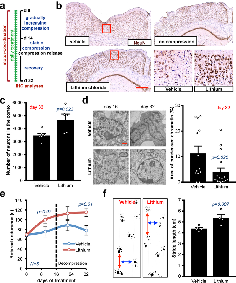
(a) Schematic of the treatments in long-term compression/decompression procedure. (b) Representative cortexes stained with anti-NeuN from a cohort of 5 mice per experimental point. Chronic lithium treatment preserves the cortex micro-anatomy after decompression. Insets are magnifications of the red squares. Scale bar: 500 μm. (c) Quantification of the NeuN+ neurons in the cortexes, decompressed or compressed and released. Data are mean ± s.e.m. (d) Ultrastructural imaging of nuclei in compressed cortexes treated with vehicle or lithium via electromicroscopy. Representative images from a cohort of 3 mice per experimental point. Scale bar: 1 μm. Quantification of the percentage of condensed chromatin area in n nuclei. Data are mean ± s.e.m. (e) Longitudinal analysis of the Rotarod endurance (index of motor coordination and balance). Data are mean of 2 consecutive days and 6 mice per group ± s.e.m. (f) Static gait test (index of locomotion); footprint analysis of the stride length at the time of highest compression (day 14). Data are mean of 3 technical replicates and n mice per group ± s.e.m. 115 steps measured for vehicle-treated mice and 62 for lithium-treated ones.