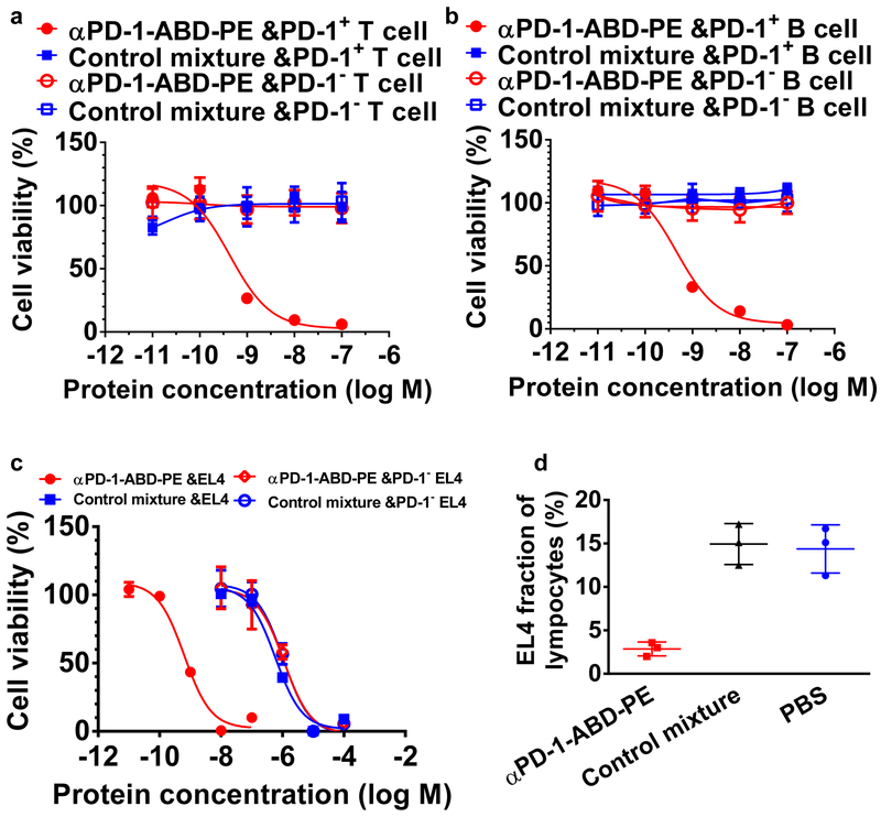
Figure 2. αPD-1-ABD-PE is selectively toxic to PD-1+ cells in vitro and in vivo.
(a) The relative viability of PD-1+ and PD-1− primary T cells after they were incubated with αPD-1-ABD-PE or a control mixture of αPD-1 and ABD-PE for 72 hours. The mean viabilities and their SDs at different concentrations of αPD-1-ABD-PE and the control mixture were shown. The viability data of PD-1+ primary T cells after the αPD-1-ABD-PE treatment were fitted to a Sigmoidal dose-response model and the IC50 was obtained through the fitting (N=6 biologically independent samples). The cells were collected from C57BL/6 mice. (b) The relative viability of PD-1+ and PD-1− primary B cells after they were incubated with αPD-1-ABD-PE or a control mixture of αPD-1 and ABD-PE for 72 hours. The mean viabilities and their SDs at different concentrations of αPD-1-ABD-PE and the control mixture were shown (N=6 biologically independent samples). The cells were collected from C57BL/6 mice. (c) The relative viability of wildtype EL4 and PD-1− EL4 cells after they were incubated with αPD-1-ABD-PE or a control mixture of αPD-1 and ABD-PE for 72 hours. The mean viabilities and their SDs at different concentrations of αPD-1-ABD-PE and the control mixture were shown and fitted to a Sigmoidal dose-response model (N=6 biologically independent samples). (d) The fractions of transferred EL4 cells among lymphocytes. These lymphocytes were collected from mice at 72 hours after these mice were treated with αPD-1-ABD-PE, a control mixture of αPD-1 and ABD-PE, or PBS. The mean faction values and their SDs are indicated (N=3 mice). All studies described in this figure were repeated twice with similar results. Data of one repeat is shown here.

