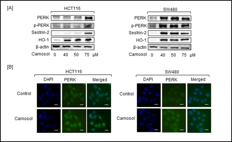
Figure 3.

Carnosol promoted the expression of PERK, p-PERK, sestrin-2 and HO-1 in treated colon cancer cells. HCT116 and SW480 cells were treated with increasing doses of carnosol for 24 hr. Cell lysates were prepared and subjected to western blot for detecting the expression of ER stress proteins and antioxidant enzymes. (A) Expression of PERK, and phosphor-PERK, sestrin-2 and HO-1 was determined by Western blot. (B) Intracellular immunofluorescent staining of PERK in carnosol-treated HCT116 and SW480 cells. Left column, DAPI-stained nuclei appear blue; middle column: FITC-stained cells appear green showing PERK expression; right column: merged pictures of DAPI and FITC. Bar scale represents 50 px.
