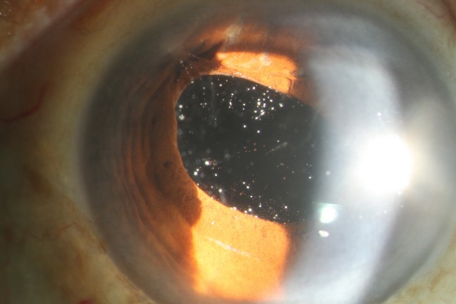
Figure 1.
Anterior segment photo of the right eye shows prolapse of vitreous in the anterior chamber with incarceration in the inner lip of the superotemporal incisional scar, causing peaking of the pupil at 10 o’clock and temporal decentration of the intraocular lens. Multiple pigments and white dots appearing like asteroid hyalosis are seen in this vitreous framework.

