Figure 6.

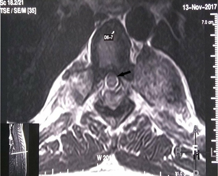
Post-radiotherapy MRI, T2 axial image at D6-7 level showing substantial reduction in the cord compression (black arrow). The inner image depicts the level of the spine at which this axial section is taken.

Post-radiotherapy MRI, T2 axial image at D6-7 level showing substantial reduction in the cord compression (black arrow). The inner image depicts the level of the spine at which this axial section is taken.