Figure 3.

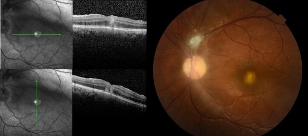
New macular yellowish lesion with moderate vitreous inflammation (right). Optical coherence tomography images showing foveal hiper-reflective lesion affecting all retinal layers (left).

New macular yellowish lesion with moderate vitreous inflammation (right). Optical coherence tomography images showing foveal hiper-reflective lesion affecting all retinal layers (left).