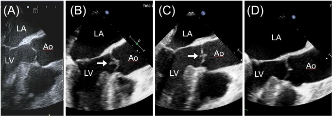
Figure 2.
Transoesophageal echocardiogram in the long-axis view on admission showing no abnormality in the aortic valve (A). A vegetation (white arrows) in the aortic valve newly developed on day 25 (B), diminished rapidly within a week of initiation of heparin infusion (C) and disappeared on day 75 (D). Ao, aorta; LA, left atrium; LV, left ventricle.

