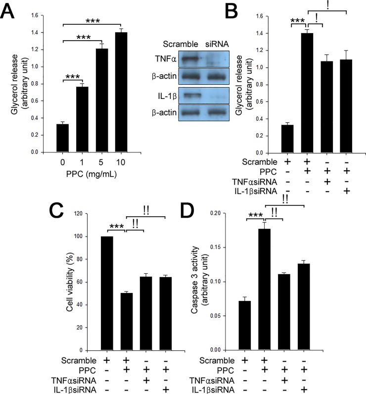
Fig 3. TNFα and IL-1β contribute to PPC-mediated lipolysis and apoptosis in adipocytes.
(A) 3T3-L1 adipocytes were treated with different concentrations (0–10 mg/mL) of PPC for 24 h. Cells were assessed by lipolysis assay. Scramble and TNFα or IL-1β siRNAs-transfected 3T3-L1 adipocytes were treated with PPC (10 mg/mL) for 24 h. Cell extracts were measured by lipolysis assay (B), MTT assay to determine cell viability (C), and caspase 3 activity (D). Means ± SEM were calculated from three independent experiments. ***P < 0.001 compared to control 3T3-L1 adipocytes. !P < 0.001 and !!P < 0.01 compared to the levels in 3T3-L1 adipocytes treated with PPC.

