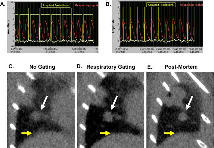
Fig 8. Prospective gating allows for synchronization of image acquisition with breathing patterns.
Micro-CT acquisition monitoring without (A) and with respiratory gating (B). Coronal images of a lung tumor are shown without gating (C), with respiratory gating (D), and post-mortem (E). Tumor is indicated by white arrows, and diaphragm is noted with yellow arrows. Representative images from triplicates are shown.

