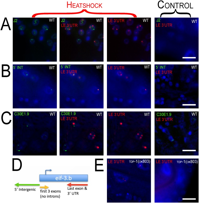
Fig 7. Fluorescence in situ Hybridization (FISH) of eif-3.B regions.
100x oil immersion images of worm hypodermal and neuronal cells. Heat shock panels are in the three columns to the left (merged channel in the middle column). Control panels show exposure from every channel (right column). Row (A) Immunohistochemistry with J2 antibody (green) along with FISH of doW01D2.8 antisense to the last exon and 3’ UTR (LE 3’ UTR) (red) of eif-3.B. dsRNA and the antisense LE 3’UTR transcript aggregate into nuclear foci with heat shock and do not appear to colocalize. Row (B) FISH of doW01D2.8 in two regions antisense to the 5’ intergenic region (5’ INT) (green) and last exon and 3’ UTR (LE 3’UTR) (red) of eif-3.B. Row (C) FISH of doW01D2.8 antisense to the last exon and 3’ UTR (LE 3’UTR) (red) of eif-3.B and sense probe of ncRNA C3DE1.9 (green). Probing of C3DE1.9 is not affected by heat shock and C3DE1.9 is not induced by heat shock. C3DE1.9 and LE 3’UTR show no overlap. (D) Diagram of eif-3.b gene with FISH probe locations and orientation. (E) Heat shock of tdp-1(ok803) induces nuclear foci from probes antisense to the last exon and 3’ UTR (LE 3’UTR) of eif-3.B (left panel) and is not visible with no heat shock (right).

