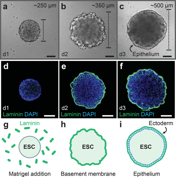
Figure 2: Development of the definitive ectoderm epithelium.
a-c, DIC images of representative aggregates on days 1, 2, and 3 of 3D culture. d-i, Following Matrigel addition on day 1, Laminin is incorporated into a basement membrane (e, f, h, i) on the surface of the aggregate. An epithelium develops on the aggregate surface by day 3. See Table 1 for a list of antibodies used for characterization. ESC, embryonic stem cells. Scale bars, 100 μm

