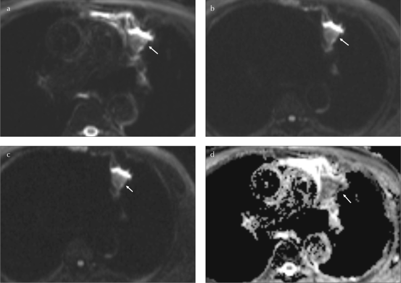
Figure 2. a–d.
A 45-year-old female without history of smoking. SPN at left lung, upper lobe, inferior lingular segment (arrows). Axial TIWI image (a).The LSR500 and LSR1000 values are 0.69 and 0.82, consequently on axial DWIs (b,c). The ADC value is 0.73 mm2/s (d). Histopathological diagnosis is squamous cell carcinoma
LSR: lesion spinal cord ratio; SPN: solitary pulmonary nodule

