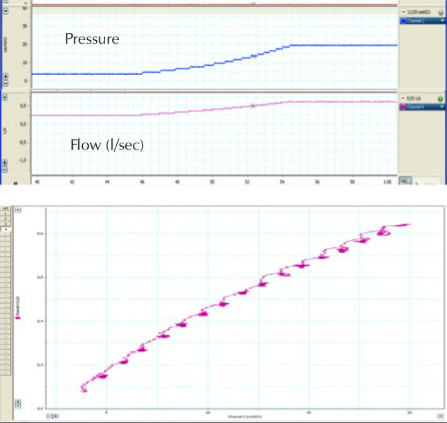
Figure 3.

Leak test performed in the laboratory with an external pneumotachograph. In the upper panel, there is a progressive increase in pressure, with the corresponding increase in flow with the distal end of the tubing occluded and an expiratory port included in the circuit. In the lower panel, a pressure-leak plot is constructed
