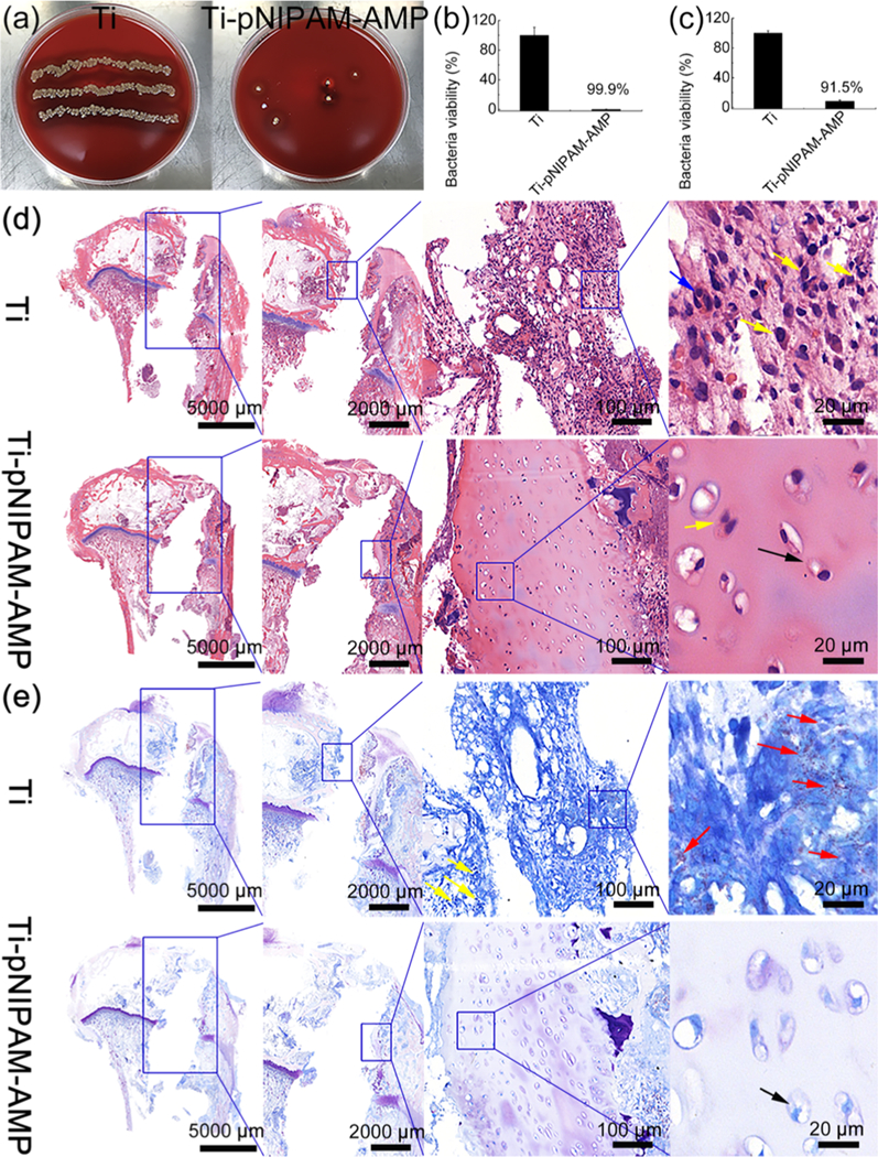
Figure 8.

In vivo characterization of antimicrobial activity and biocompatibility of samples after implanted in rabbit tibiae for 7 d. Before implantation, the samples were incubated with bacteria at RT. (a) Images of the Petri dishes showing the presence of bacteria (yellow spots) after the implanted samples (Left: Ti, Right: Ti-pNIPAM-AMP) were rolled over the blood agar. (b,c) Antimicrobial activity of the surfaces of different samples (b) and the tissues surrounding the corresponding samples (c). These data suggest that Ti-pNIPAM-AMP is more antimicrobial than Ti, leading to much fewer bacteria on both itself and its surrounding tissues. (d,e) Histological images of H&E (d) and Giemsa (e) staining. The blue and yellow arrows denote osteoclasts and inflammatory cells, respectively. The red arrows denote the bacteria observed in the tissue. The black arrows denote the live bone cells. These data suggest that Ti-pNIPAM-AMP presented an antimicrobial activity at RT and increased biocompatibility in vivo.
