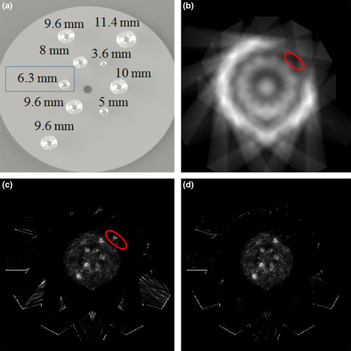
Figure 7.

(a) Illustration of the sizes and distribution of the spherical tumor inserts in the cylindrical phantom; (b) Sensitivity image of the prototype POC‐PET system; (c) Image of a cylindrical phantom containing nine spherical tumors with 8:1 tumor‐to‐background radioactivity concentration, reconstructed without TOF information; (d) Image reconstructed with TOF information. [Color figure can be viewed at wileyonlinelibrary.com]
