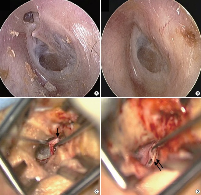
Fig. 2.
Microscopic approach for attic cholesteatoma in patient who suffered from intermittent otorrhea and otalgia (case 16 in Table 1). (A) Attic destruction was observed on preoperative examination of the left tympanic membrane. (B) Postoperative examination shows the tympanic membrane with clear attic area. Intraoperative findings show the elevated tympanomeatal flap (arrow) after Lempert endaural incision II and Lempert I incision (C), and inserted cartilage for reconstruction of the attic area (double arrows, D).

