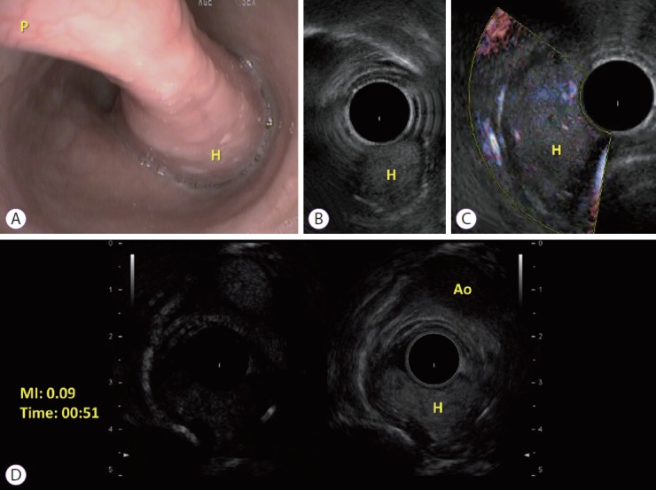
Fig. 2.

(A) Endoscopic view of the esophageal fibrovascular polyp. Long thin peduncle (P) and the head (H) of the polyp; (B) Ultrasound endoscopy imaging of the head (H) of the polyp; (C) Idem B with Fine doppler; (D) Contrast-enhanced endoscopic ultrasound of the head (H) of the polyp at the same plane of the aorta (Ao), with low mechanical index (MI).
