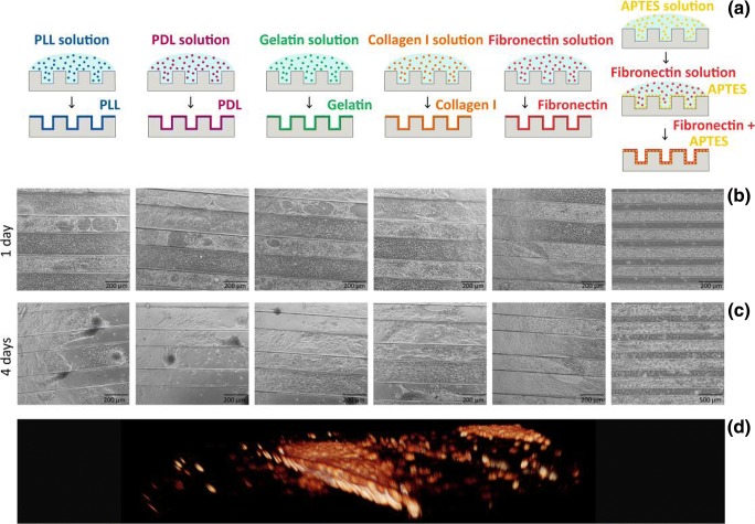
Fig. 2.
Establishment of microgroove culture system for ESPCs. a The PDMS substrates are functionalised with different coating strategies. b-c Phase-contrast images of ESPCs cultured on substrates with different coating strategies at (b) 1 DIV and (c) 4 DIV. Cell detachment and aggregation is visible at day 4 except for the APTES+fibronectin strategy. d Volumetric reconstruction of a confocal stack shows the 3D shape of the condensations in the microgrooves

