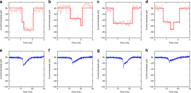
Fig. 6. Sample transient current traces zoomed in from Fig. 5b, c.
For the 12 nm pore in (a–d), most peaks look almost symmetric with different translocation modes, such as linear, double local folded, single local folded or fully folded fragments of DNA molecules, whereas peaks for the 6 nm pore in (e–h) are asymmetric, which we attribute to the larger entropic barrier associated with the smaller pore; the DNA molecule resides at the tip of the pore for a certain duration in a coiled state prior to overcoming the entropic barrier to then thread into the nanopore in a linear fashion

