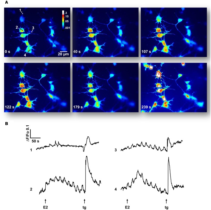
Figure 2.
17β-estradiol (E2) induces Ca2+ oscillations. (A) Hypothalamic neurons were loaded with Cal-520 AM for 30 min at 37°C, maintained in a Ca2+-containing buffer (Ca2+-HBSS) and changes in cytosolic Ca2+ concentration were measured using confocal microscopy [Olympus IX81 inverted microscope equipped with a Disk Spinning Unit (DSU)]. Time series of Cal-520 pseudocolor images is shown, before and after the addition of E2 and thapsigargin (tg). Pseudocolor scale bar: 0.3–0.001 arbitrary units. Length scale bar: 20 μm. (B) Representative Ca2+ traces [regions of interest labeled 1–4 in (A)] plotted as changes over time in fluorescence intensity of the indicator (ΔF) respect to resting values (Fo). Arrows indicate the addition time of E2 (30 s) and tg (3 min). Data are from one representative experiment out of six independent experiments.

