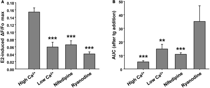
Figure 3.
Ryanodine, external Ca2+ and nifedipine modulate E2-induced Ca2+ increase. (A) Mean values of maximal ΔF/Fo for Ca2+ mobilized by 17β-estradiol (E2) in hypothalamic neurons recorded either in the presence or absence of extracellular Ca2+ (Ca2+-HBSS/EGTA-containing buffer, named High Ca2+/Low Ca2+), or after a pre-incubation period (1 h) with ryanodine or nifedipine. ANOVA F(3,41) = 22.94; p ≤ 0.001. LSDs test indicated ***p < 0.001 vs. Ca2+-HBSS. (B) Mean values of integrated area under ΔF/Fo curve (AUC) after thapsigargin (tg) addition during Ca2+ imaging [same conditions as (A)]. ANOVA F(3,26) = 9.18; p ≤ 0.001. LSDs test indicated ***p < 0.001 and **p = 0.01 vs. ryanodine. Bars represent mean ± SEM; n = 4–6 different cultures.

