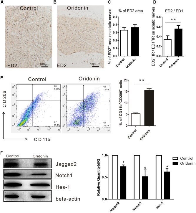
FIGURE 5.

Oridonin increased proportion of anti-inflammatory macrophages in EAN rats through suppression of Notch pathway. Rats were treated therapeutically with Oridonin and sacrificed on Day 15; sciatic nerves were taken for immunohistochemical staining. By staining, anti-inflammatory macrophages were identified by expression of CD163, which is detected by ED2 antibody. Representative microphotos show ED2+ cells in sciatic nerves of EAN rats treated by Oridonin (B) in comparison to the control (A). In the control group, much more infiltrates could be observed, but CD163 expression was not significantly different from the treatment group, where much less accumulated immune cells could be seen. (C,D): The bar graphs show no difference in CD163 IR levels between control and Oridonin groups, but the proportions of ED2+ over ED1+ IR areas in sciatic nerves were significantly increased in comparison to controls. Further, the frequency of CD206+CD11b+ cells from spleen were tested by flow cytometry. (E) Spleen mononuclear cells (MNCs) were isolated from the control and Oridonin treated EAN rats at day 15 post-immunization and examined by flow cytometry. Percentage of anti-inflammatory macrophages (CD206+CD11b+ cells) in spleen MNCs was significantly increased in Oridonin treated rats compared to the control group. (F) On day 15, sciatic nerves of each rat were harvested for Western blotting. Relative expression of Jagged-2, Notch1 and Hes-1 in Oridonin-treated groups was significantly lower compared to the control group. ∗p < 0.05, ∗∗p < 0.01 compared to the control group, n = 6.
