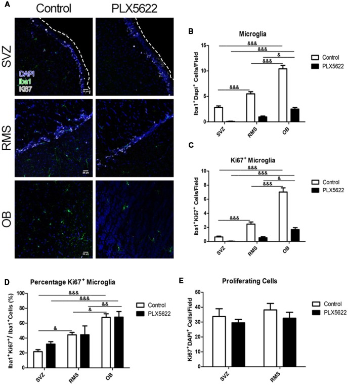
Figure 5.
Treatment with PLX5622 has no effect on microglia proliferation. Representative images of the SVZ, RMS, and OB from 14d of PLX5622 treatment (A), with the dashed line separating the SVZ from the lateral ventricle. DAPI in blue, Iba1 in green, Ki67 in gray. Quantification of microglia ablation shown in (B, F(2,15) = 55.46, p < 0.0001). Ki67+Iba1+ cells were counted (C, F(2,15) = 64.52, p < 0.0001) and normalized (D, F(2,15) = 12.77, p = 0.0006). Similar to the BrdU data, the OB contained more Ki67+ microglia than the RMS and SVZ in both control and PLX5622 treated mice. Total Ki67+ cells from the SVZ and RMS were quantified in (E, F(2,15) = 0.11, p = 0.74). & = comparison between regions within treatment, & = p < 0.5; && = p < 0.01; &&& = p < 0.001. n = 6 for all groups.

