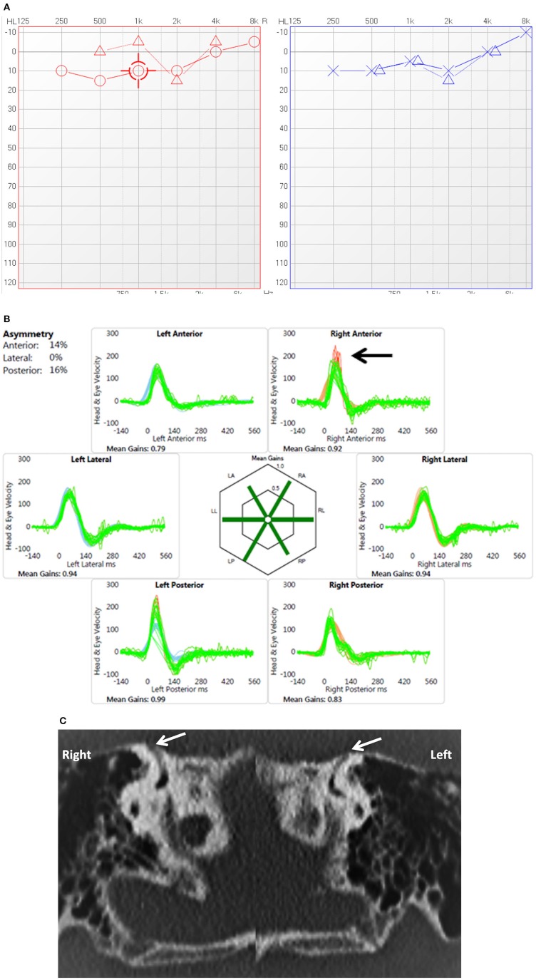
Figure 3.
(A) PTA in child presenting with conductive dysacusis (child 7). (B) Video head impulse test in a child with covert saccades in the right superior semicircular canal (arrow). (C) Coronal reconstruction of High resolution CT scan shows dehiscence of the superior semicircular canal on both sides (arrows).

