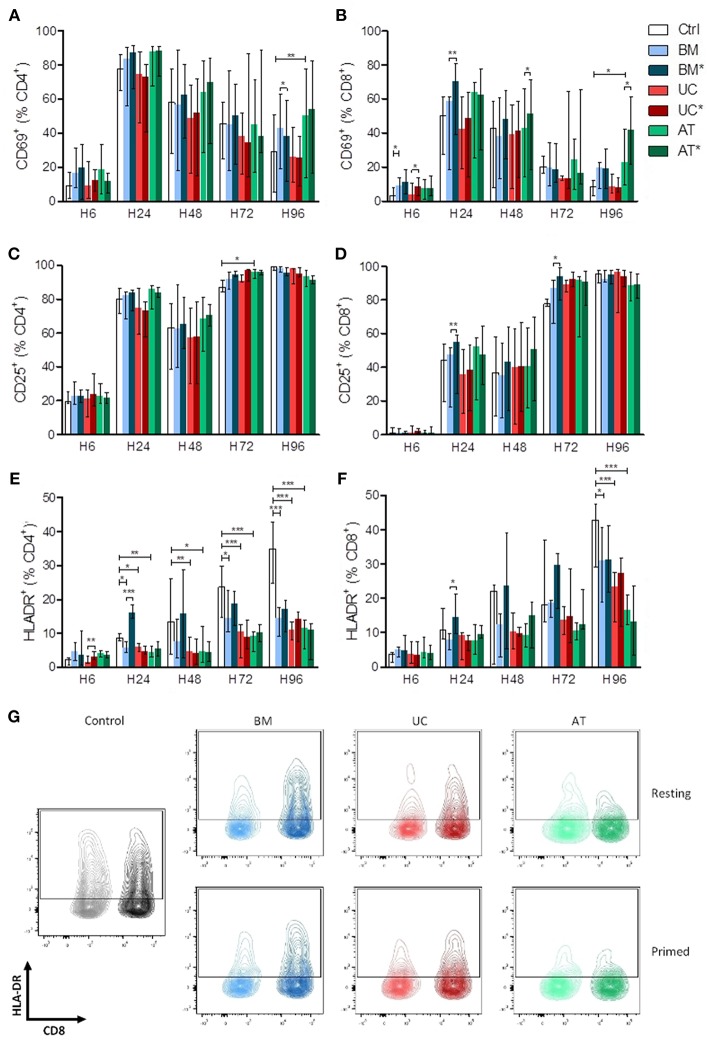
Figure 2.
Lymphocyte activation (measured by CD69, CD25, and HLA-DR expression) in co-culture with MSC. PBMCs were cultured with or without MSCs in the presence of anti-CD3/CD28 microbeads for 4 days, at a MSC/PBMC ratio of 1/10. For inflammatory stimulation, MSCs were incubated with IFNγ 10 ng/ml and TNFα 15 ng/ml during 40 h, prior to harvest (BM*, AT*, and UC*). Expression of (A,B) CD69, (C,D) CD25, and (E,F) HLA-DR on CD4+ and CD8+ lymphocytes was analyzed after 6, 24, 48, 72, and 96 h by FACS. (G) Representative plots of HLA-DR expression at H96 in CD4+ and CD8+ lymphocytes. Data are presented as median with range. Differences between control, and BM, AT, or UC groups are calculated with repeated measure ANOVA with Dunnett's post-hoc procedures (only results of Dunnett's post-hoc tests are represented). Differences between resting and primed MSC groups were calculated with paired t-test (*p < 0.05, **p < 0.01; ***p < 0.001).

