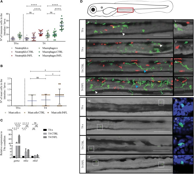
Figure 1.
Neutrophils, macrophages, and mast cells increase their presence in the gut during the inflammatory process triggered in the naïve feeding model. The number of neutrophils/macrophages (A), and mast cells (B) was quantified under different conditions (n, naïve; CTRL, control; INFL, inflammation) and time points (T0: 5 dpf; T4: 9 dpf). (C) Relative mRNA levels of gzma, nkla, and nkld were analyzed. Data were normalized against rpl13a and compared to T4 naïve condition (dotted line). (D) Scheme of a lateral view of a 9 dpf larva with the zone of the intestine used to quantify delimited by the red rectangle. (E–H) Lateral view of the intestine of a Tg(lysC:DsRed)xTg(mpeg1:Dendra2) larva showing fluorescently labeled neutrophils (red) and macrophages (green). (M–P) Lateral view of the intestine showing mast cells labeled by anti-tryptase immunohistochemistry. Zoom of macrophages and neutrophils (I–L) and mast cells (Q–T). *p < 0.05; ***p < 0.001; ****p < 0.0001. Scale bar 200μm. Experiments were done at least in three biological replicates with 20 individuals per condition.

