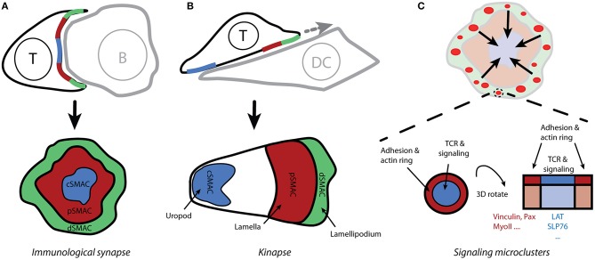
Figure 2.
Spatio-temporal organization of T-cell activation. (A) The contact site between T cells and antigen-presenting cells (which form conjugates) is called the immunological synapse due to its similarity to neuronal synapses. Live-cell confocal microscopy reveals the supramolecular organization of molecules within the immunological synapse, with the receptors and effector molecules accumulating in the center of the mature synapse (cSMAC) and adhesion molecules forming a peripheral ring (pSMAC; i.e., the bull's eye model). (B) Motile T cells form asymmetric kinapses instead of stable and symmetric synapses. Kinapses are similar to motile fibroblast cells with lamellipodium (dSMAC), lamella (pSMAC), and uropod (cSMAC). (C) Total internal-reflection fluorescence microscopy reveals that T-cell signaling is initiated in small microclusters that are assembled upon antigenic stimulation in actin-rich distal regions of the immunological synapse (A; dSMAC). Small rings of adhesion molecules and actin surround TCR microclusters and could serve to stabilize those microclusters.

