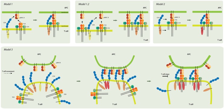
Figure 3.
Models of CD4 role in T-cell activation. Model 1 (upper left panel). TCR and CD4 independently interact with pMHC, and the coreceptor stabilizes the receptor-ligand complex. Inactive Lck (yellow) becomes partially activated (light pink). Model 1.2 (upper middle). In this pseudo-dimer model, two TCRs interact with two pMHCs. One MHC always contains an agonist peptide (dark brown), but the other can have a non-stimulating (self) peptide (light brown) instead. The role of CD4 is to crosslink the two receptor-ligand pairs and to thus accelerate signaling. Model 2 (upper right). CD4 delivers the Lck kinase to the formed TCR-pMHC super-complex that has the antigenic peptide. Lck is fully activated (dark pink) by binding to ZAP-70 (not shown) and accommodating open conformation. Model 3 (lower panel). CD4 accumulates at the tips of T-cell microvilli. Independent of TCR, the CD4 that is on microvilli interacts with the pMHC that is assembled on similar membranous protrusions of antigen-presenting cells (APC). This multivalent interaction drives putative changes in the tips of the microvilli and activates Lck; it thus provides T cells and their TCRs with high sensitivity to antigenic peptides. The presented models are not mutually exclusive. Color-coding of CD4, TCR/CD3 complex, and MHCII as in Figure 1.

