Figure 2.

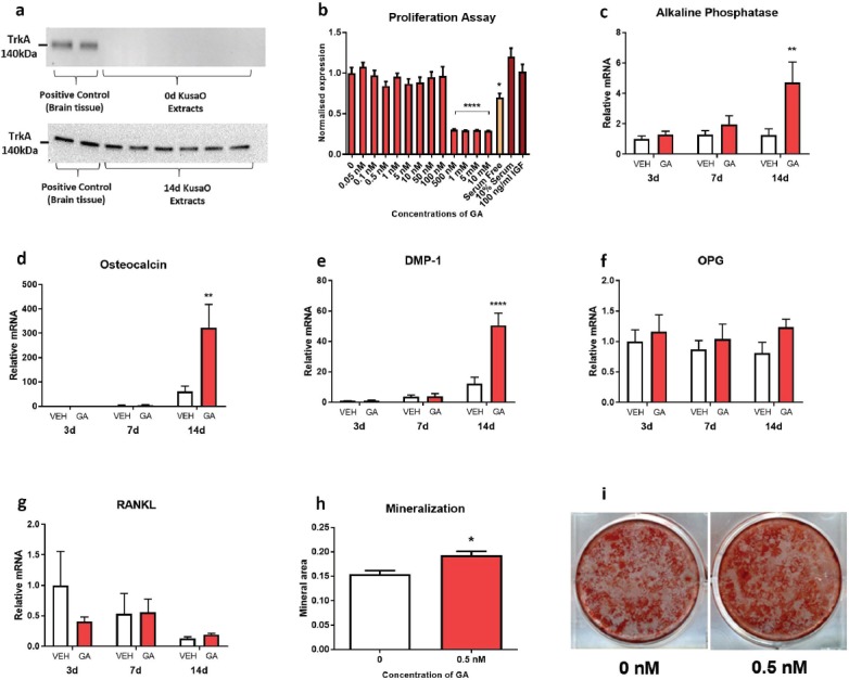
TrkA expression and the effects of GA treatment in the mesenchymal stem cell line, Kusa O. Western blotting (a) revealed that differentiated but not undifferentiated KusaO cells expressed TrkA. GA did not increase proliferation after 72 h of treatment, however, GA was cytotoxic at concentrations ≥500 nM (b; ****p<0.0001). Gene expression markers of mature osteoblasts (c,d), early osteocytes (e), and osteoclast formation (f,g), were analyzed at 3-, 7-, and 14-days of differentiation. GA treatment increased alkaline phosphatase (c; **p<0.01), osteocalcin (d; **p<0.01), and DMP-1 (e; ****p<0.0001) gene expression of Kusa O’s at 14 days compared to vehicle. Time or GA had no effect on OPG or RANKL expression (f,g). Kusa O cells formed mineral after 21 days of incubation. GA increased mineralization of Kusa O cells compared to vehicle (h,i; *p<0.05). GA, gambogic amide; DMP-1, dentin matrix acidic phosphoprotein 1; OPG, osteoprotegrin; RANKL, receptor activator of nuclear factor kappa-B ligand. Bars are mean ± SEM, n=6-8/group.
