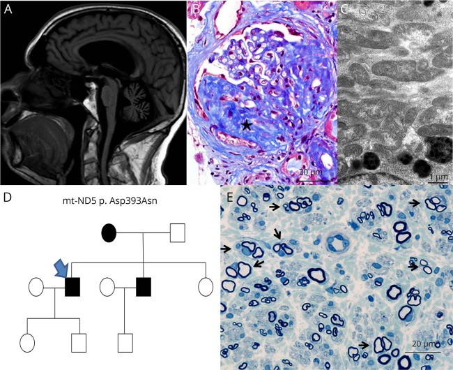
Figure. ND5 and MCARNE phenotype.
(A) Brain MRI sagittal T1 FLAIR showing moderate generalized cerebral and severe cerebellar atrophy without evidence of previous strokes. (B) Kidney biopsy revealed a subset of glomeruli with segmental glomerulosclerosis (marked star) as is seen here at ×400 magnification with Masson trichrome stain. (C) Morphologically, the mitochondria were well within the range of normal variation as is seen in electron micrograph at ×13,000 magnification. (D) Pedigree chart; index case (arrow). (E) Methylene blue epoxy section of the patient's brother's sural nerve biopsy at ×500 magnification showing clusters of regenerating myelinated axons surrounded by concentric Schwann cell processes (arrows) with onion bulb–like formations.

