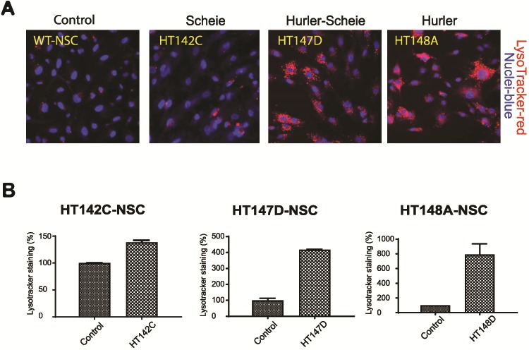
Figure 2.

Validation of lysosome accumulation. (A) Variable degree of lysosomal accumulation displayed in patient NSCs in the presence of serum. LysoTracker® staining in MPS I-NSCs in presence of serum exhibit enlarged lysosomes. In comparison to the control line, Hurler cells displayed abundant lysosomal accumulation, whereas H/S was intermediate and Scheie weaker. (B) Quantitative In Cell analysis correlation with enlarged lysosomal phenotypic staining visualized in the NSC cells.
