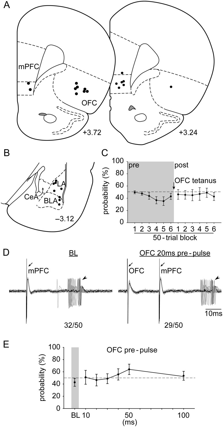
Figure 5.
The placements of (A) all the stimulation electrodes in the mPFC and the OFC and (B) the distribution of all the neurons recorded (+3.72, +3.24, and −3.12; AP distance [mm] to bregma). (C) OFC tetanus did not change evoked probability of the amygdala neuron response to mPFC stimulation. (D) Electrophysiological recording of an amygdala neuron that responded to mPFC stimulation after OFC tetanus. (E) Stimulation of the OFC failed to produce an inhibitory gating over the mPFC–amygdala pathway following OFC tetanus. Abbreviations refer to Figures 1 and 3.

