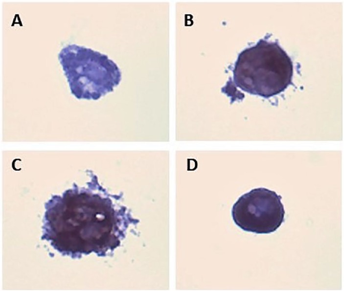
Figure 7.
Immunohistochemistry (IHC) of 3T3-L1 cells grown in three-dimensional (3D) agarose. Cultures were processed for routine light microscopy, embedded in paraffin and sectioned at 5 µm. (A) Cell (2.5 weeks in culture) represents a negative control (no primary PPARγ antibody) prior to secondary reaction with HRP and DAB chromogen. (B) Cells grown for 0.5 weeks stained with antibody for PPARγ. Evidence of PPARγ demonstrated by the DAB chromogen (brown) is indicative of adipogenesis. (C) Cells grown for 1.5 weeks. (D) Cells grown for 2.5 weeks. Compared with control, all cells are positive for PPARγ and demonstrate adipogenesis. Slides were stained with hematoxylin for 30 seconds to make visible in the microscope. DAB indicates 3,3′-diaminobenzidine; HRP, horseradish peroxidase; PPARγ, peroxisome proliferator-activated receptor γ.

