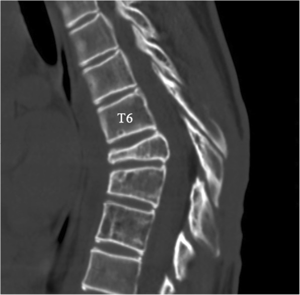
Fig. 10.

Thoracic CT at 2-year follow-up. Axial and sagittal CT scans show bilateral pleural thickening and minimal pleural effusion; changes in the thoracic spine and osteolytic rib stopped. (The white arrow indicates the destructed vertebrae, the red arrow indicates the osteolytic rib, and the circle indicates pleural effusion)
