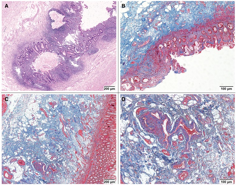
Figure 1.
Morphologic features of radiation-induced bowel injury. (A) Crypt disarrangement and mucosal inflammatory infiltrate (Hematoxylin & Eosin staining, 40× field; histopathological score, 2 for both). (B) Mucosal erosions, fibrosis of the lamina propria and fibrosis of submucosa (Masson’s trichrome staining, 100× field; histopathological score, 1, 1 and 2, respectively). (C) Fibrosis of submucosa (Masson’s trichrome staining, 40× field; histopathological score, 2). (D) Sclerosis of submucosal vessels (Masson’s trichrome staining, 100× field; histopathological score, 2).

