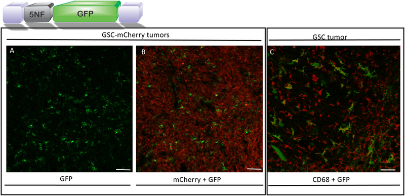
Figure 3. Systemic injection of exo-AAV9–5NF-GFP transduces tumor-associated myeloid derived cells in human glioma stem-like cell tumors in mice.
exo-AAV9–5NF-GFP transduction of brain tumor stromal cells after intravenous injection. Nude mice bearing intrastriatal human GSC tumors were injected i.v. with exo-AAV9–5NF-GFP (vector schematic shown above; purple blocks, AAV inverted terminal repeats; 5NF, NF-κB inducible promoter). Mice were sacrificed at the humane endpoint, tumors sectioned, and immunostained for GFP. (A) GFP expression in tumor stromal cells. (B) Merge of GFP signal with tumor cell-expressed mCherry to show that the transduced cells are within the tumor mass. Scale bar=100 μm. (C) exo-AAV9–5NF-GFP primarily transduces CD68+ myeloid-derived cells within human GBM tumor (no mCherry expression in this tumor. Confocal imaging of human GSC tumor-bearing mice injected i.v. with exo-AAV9-NF-GFP. Tumor sections were stained with myeloid-derived cell marker CD68 (red) and co-localization with GFP was readily detected. Scale bar= 50 μm.

