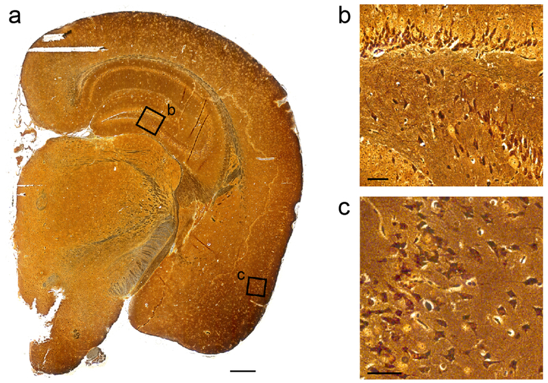
Fig. 3. Tg(SNCA*A53T+/+)Nbm mice develop silver-positive aggregates.
One slide from four MSA-inoculated Tg(SNCA*A53T+/+)Nbm mice with phosphorylated α-synuclein neuropathology were analyzed for the presence of silver-positive aggregates. Representative full section (a) shows magnified regions of the hippocampus (b) and piriform cortex (c) in black boxes, which contain labeled aggregates. Scale bar in (a), 500 μm. Scale bars in (b, c), 50 μm.

