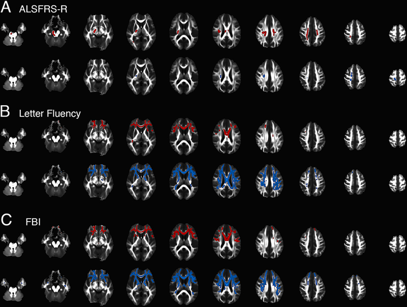
Figure 3.

Correlation between FA (red) or MD (blue) in whole-brain white matter analysis and scores on clinical scales at the baseline visit in 28 subjects with C9orf72 mutations. A) Regions of FA (top row, red) correlated with the ALSFRS-R are largely confined to portions of the CST from subcortical white matter to pons. There are small regions of correlated MD in the right CST (blue, bottom row). B) Letter fluency scores are correlated with FA (top row, red) and MD (bottom row, blue) predominantly in frontal white matter and C) Frontal Behavioural Inventory scores are correlated with FA (top row, red) and MD (bottom row, blue) in similar frontal regions.
