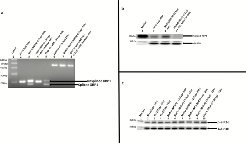
Figure 2.
A, The mRNA of XBP1 is spliced by the endonuclease activity of IRE1α. Total RNA was extracted from C57epi cells cultured in various experimental conditions. Primer pair (forward: 5’-GAACCAGGAGTTAAGAACACG -3’; reverse: 5’AGGCAACAGTGTCAGAGTCC -3’) was used in PCR amplification. The DNA template was the complementary DNA reverse transcribed from the RNA extracted from each culture condition. The PCR product from each culture condition was ran on 2% agarose gel and stained with ethidium bromide. Lane 1: molecular weight marker. Lane 2: noninfected C57epi cells cultured for 48 hours showing the presence of unspliced 205-bp band used as negative control. Lane 3: C57epi cells infected with C. muridarum (MoPn) at MOI 5 for 48 hours showing the presence of unspliced 205-bp band as well as spliced 179-bp band. Lane 4: C57epi cells infected with MoPn at MOI 5 in the presence of inhibitor of IRE1α RNase activity for 48 hours showing the presence of unspliced 205-bp band. Lane 5: Noninfected C57epi cells treated with thapsigargin (chemical inducer of UPR) cultured for 48 hours showing the presence of unspliced 205-bp band as well as spliced 179-bp band used as positive control. Lanes 6–8: PCR amplification of GAPDH gene products used as loading control for noninfected C57epi cells infected with MoPn at MOI 5 and C57epi cells infected with MoPn at MOI 5 in the presence of inhibitor of IRE1α RNase activity for 48 hours showing the presence of unspliced 205-bp band. B, C57epi cells infected with C. muridarum induced the upregulation of XBP1 protein and inhibition of IRE1α RNase activity results in the downregulation of XBP1. Ten μg total protein from C57epi cells uninfected/infected with C. muridarum for 48 hours and treated/nontreated with inhibitor of IRE1α RNase activity were prepared for Western blot analysis. Blot was probed with primary antibody against spliced XBP1. Lane 1: protein molecular weight marker. Lane 2 sample from noninfected C57epi cells at 48 hours. Lane 3: sample from C57epi cells infected with MoPn at MOI 5 for 48 hours. Lane 4: sample from C57epi cells infected with MoPn at MOI 5 in the presence of inhibitor of IRE1α RNase activity for 48 hours. C, Western blot of eIF2α phosphorylation during Chlamydia infection. Ten μg total protein from C57epi cells infected with C. muridarum (MoPn) for 24, 48, and 72 hours were prepared for Western blot analysis. Blot was probed with primary antibody against phosphorylated eIF2α. Lane 1: protein molecular weight marker. Lanes 2–4: samples from noninfected C57epi cells at 24, 48, and 72 hours. Lanes 5–7: samples from C57epi cells infected with C. muridarum (MOI 1) at 24, 48, and 72 hours. Lanes 8–10: samples from C57epi cells infected with C. muridarum (MOI 5) at 24, 48, and 72 hours. Abbreviations: bp, base pairs; C57epi cells, mouse oviduct epithelial cells; eIF2α, eukaryotic initiation factor 2-α; GAPDH, glyceraldehyde-3-phosphate dehydrogenase; IRE1α, inositol-requiring enzyme-1α; MOI, multiplicity of infection; MoPn, mouse pneumonitis; mRNA, messenger RNA; NI, noninfected; PCR, polymerase chain reaction; RNase, ribonuclease; Thap, thapsigargin; XBP1, X-box binding protein 1.

