
FIGURE 2.
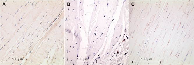
Histological sections from samples of fascia. ASMA positive stress fiber bundles—used as a marker for MFBs—were stained in dark brown, while cell nuclei were stained in dark blue. (A) Section from rat lumbar fascia. (B) Section from human fascia lata with a very low MFB density. (C) Section from human lumbar fascia. Microscopic inspection shows obvious differences in ASMA density.