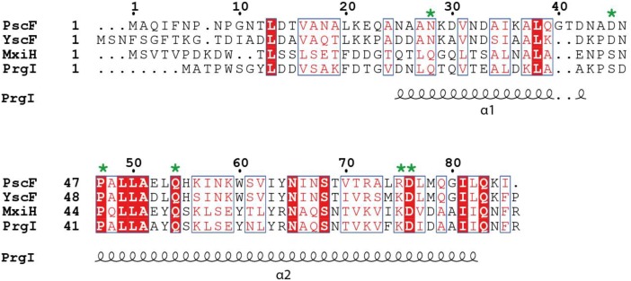
FIGURE 1.
Sequence alignment of needle proteins from different bacteria. PscF from Pseudomonas aeruginosa, YscF from Yersinia sp., MxiH from Shigella sp., PrgI from Salmonella sp. The residues mutated in this study are indicated with a green asterisk. Numbers refer to the PscF sequence. Secondary structure elements from the PrgI structure are indicated underneath the sequence alignment. The figure was generated with ESPript, using the new ENDscript server (Robert and Gouet, 2014). Conserved and similar residues are shown in red and blue boxes, respectively.

