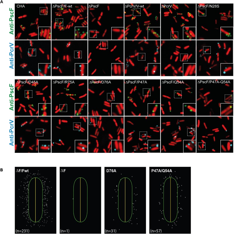
FIGURE 4.
Localization of PscF-needles on the surface of Pseudomonas aeruginosa. (A) P. aeruginosa CHA strain isolated on cystic fibrosis patient depleted for the pscF gene (ΔpscF) and complemented with pIApG-pscF constructs were grown in T3SS-inducing conditions. PscF needles were visualized by immuno fluorescence on fixed bacteria using anti-PscF (in green) or anti-PcrV (in cyan). SYTO24 was used to visualize bacteria (in red). As a negative controls we used P. aeruginosa ΔF and ΔV, while ΔF/Fwt and ΔV/Vwt were used as a positive control. Only few PscF and PcrV spots were visible on P. aeruginosa strains carrying a PscF-D76A or a PscF-P47A/Q54A. (B) MicrobeJ reconstitution of PscF distribution around the bacteria. n = number of spots in the image.

